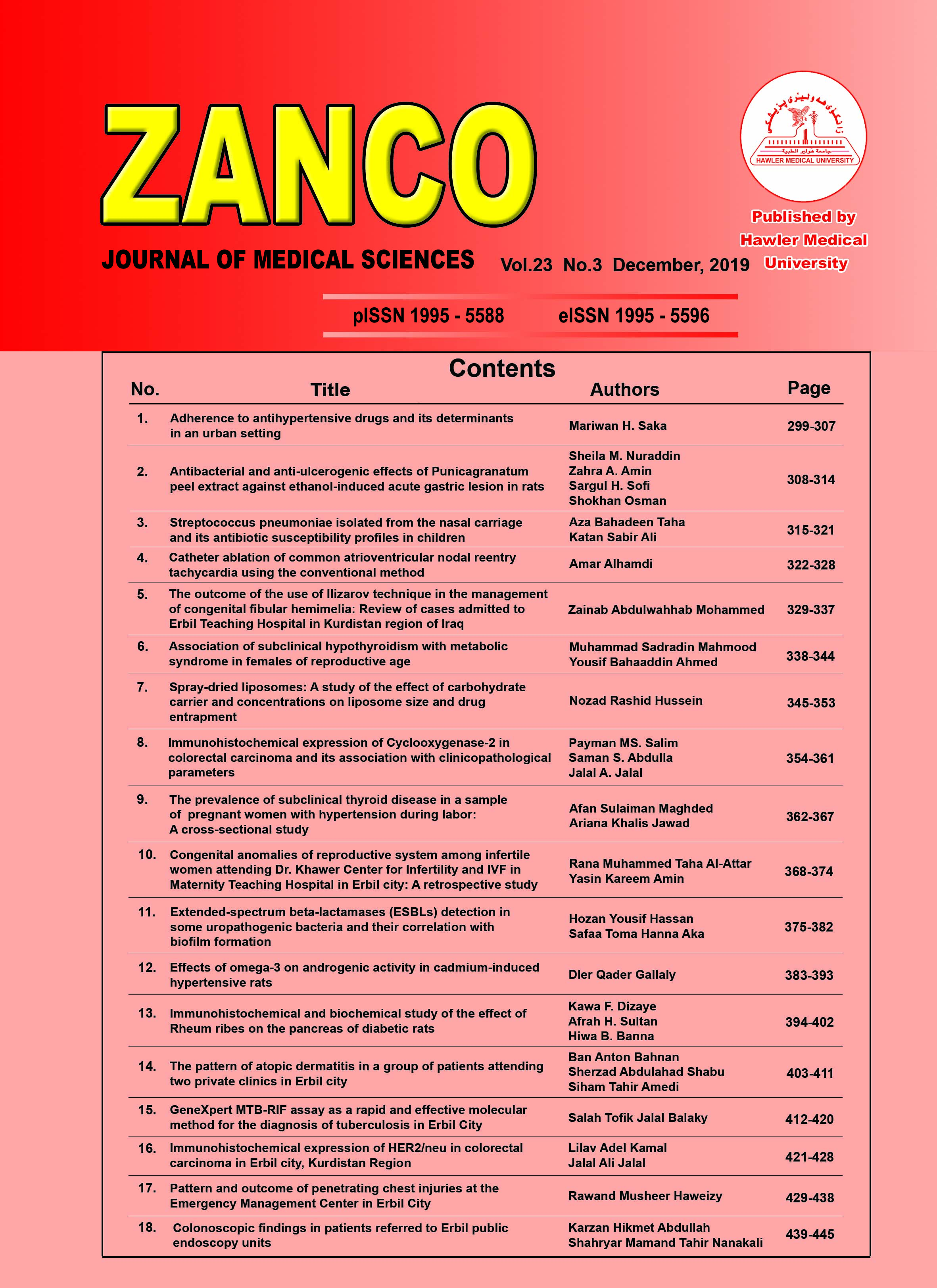
Congenital anomalies of reproductive system among infertile women attending Dr. Khawer Center for Infertility and IVF in Maternity Teaching Hospital in Erbil city: A retrospective study

This work is licensed under a Creative Commons Attribution-NonCommercial-ShareAlike 4.0 International License.
- Articles
- Submited: May 12, 2020
-
Published: December 1, 2019
Abstract
Background and objective: Congenital anomalies of the female genital tract is a known cause for infertility and recurrent pregnancy losses. This study aimed to evaluate the pattern and prevalence of each type of congenital malformation of the reproductive tract among infertile women attending the IVF center in the Maternity Teaching Hospital in Erbil city.
Methods: A retrospective study was conducted in Center of Dr. Khawer for Infertility and IVF in the Maternity Teaching Hospital in Erbil city. Data of five years from the 1st January 2010 to the 1st January 2015 were collected from patient files, such as age, sex, residence, type of infertility, type of anomaly, duration of infertility, Karyotype, Intervention, Success rate of intervention, cycle, parity, history of abortions, history of preterm deliveries, and history of ectopic pregnancy.
Results: The prevalence of congenital reproductive system anomaly was 4.1% among infertile women. The sub-septate uterus was the most common type (23.24%) anomalies. No significant association was found between the types of the anomaly and the type of infertility. The genetic factor was found to has a significant role in developing such a condition. Success rate after management significantly raised to 61.1% with sub-septate uterus (91.3%) bicornuate/ partial (85.7%), and septate/ complete (72.2%).
Conclusion: One of the causative factors of female infertility is congenital anomaly. Most types have a good prognosis in achieving pregnancy after proper management.
Metrics
References
- Hassan MA, Lavery SA, Trew GH. Congenital uterine anomalies and their impact on fertility. Women’s Health 2010; 6(3):443–61.
- Iverson RE, DeCherney AH, Laufer MR. Clinical manifestations and diagnosis of congenital anomalies of the uterus. Up-To-Date 2012; 20(3):1–4.
- Keith L, Moore KL. The developing human: clinically oriented embryology. 10thed. Philadelphia, PA: Saunders; 2016. P. 51–267.
- Sadler TW. Urogenital system, Langman's Medical Embryology. 13th ed. Philadelphia, PA: Lippincott Williams & Wilkins; 2015. P. 261–76.
- DeSilva M, Munoz FM, Mcmillan M, Kawai AT, Marshall H, Macartney KK, et al. Congenital anomalies: Case definition and guidelines for data collection, analysis, and presentation of immunization safety data. Vaccine 2016; 34(49):6015.
- Toufaily MH, Westgate MN, Lin AE, Holmes LB. Causes of congenital malformations. Birth Defects Res 2018; 110(2):87–91.
- Grimbizis GF, Gordts S, Di Spiezio, Sardo A, Brucker S, De Angelis C, et al. The ESHRE/ESGE consensus on the classification of female genital tract congenital anomalies. Hum Reprod Open 2013; 28(8):44–2032.
- Acién P, Acién MI. The history of female genital tract malformation classifications and proposal of an updated system. Hum Reprod Update 2011; 17(5):693–705.
- Heinonen PK. Renal tract malformations associated with Müllerian duct anomalies. Clin Obstet Gynecol Reprod Med 2018; 4(1):3–5.
- Arbuckle JL, Hoover KH. Development of the Female Reproductive Tract and Associated Anomalies. Curr Treat Options Pediatr 2016; 2(3):131–42.
- Hua M, Odibo AO, Longman RE, Macones GA, Roehl KA, Cahill AG. Congenital uterine anomalies and adverse pregnancy outcomes. Am J Clin Exp Obstet Gynecol 2011; 205(6): 558.e1–5.
- Woelfer B, Salim R, Banerjee S, Elson J, Regan L, Jurkovic D. Reproductive outcomes in women with congenital uterine anomalies detected by three-dimensional ultrasound screening. Obstet Gynecol 2001; 98(6):1099–103.
- Acién P, Acién MI. The history of female genital tract malformation classifications and proposal of an updated system. Hum Reprod Update 2011; 17(5):693–705.
- Grimbizis GF, Gordts S, Di SpiezioSardo A, Brucker S, De Angelis C, Gergolet M, et al. The ESHRE/ESGE consensus on the classification of female genital tract congenital anomalies. Hum Reprod 2013; 28(8):2032–44.
- Oyetunji I, Aikhuele H, Adejuwon O, Afolabi BM. Pattern of Congenital Uterine Anomalies among Infertile Women With and Without Recurrent Miscarriages, in Southwest Nigeria. Crit Care Obst Gyne 2015; 1(1):7.
- Robbins JB, Broadwell C, Chow LC, Parry JP, Sadowski EA. Müllerian duct anomalies: embryological development, classification, and MRI assessment. J Magn Reson Imaging 2015; 41(1):1–2.
- Al-Allaf LI. Frequency of Congenital Anatomical Uterine Abnormalities in a Group of Infertile Women in Mosul city. IJCM 2009; 22(4):238–46.
- Chan YY, Jayaprakasan K, Zamora J, Thornton JG, Raine-Fenning N, Coomarasamy A. The prevalence of congenital uterine anomalies in unselected and high-risk populations: a systematic review. Hum Reprod Update 2011; 17(6):761–71.
- Saravelos SH, Cocksedge KA, Li TC. Prevalence and diagnosis of congenital uterine anomalies in women with reproductive failure: a critical appraisal. Hum Reprod Update 2008; 14(5):415–29.
- Ajayi A, Ajayi V, Biobaku O, Oyetunji I, Aikhuele H, Adejuwon O, et al. pattern of congenital uterine anomalies among infertile women with and without recurrent miscarriages, in Southwest Nigeria. Critical Care 2015; 1(1):7.
- Ribeiro SC, Tormena RA, Peterson TV, Gonzáles MD, Serrano PG, Almeida JA, Baracat EC. Müllerian duct anomalies: review of current management. Sao Paulo Medical Journal 2009; 127(2):92–6.
- Tiker F, Yildirim SV, Barutcu O, Bağiş T. Familial müllerian agenesis. The Turkish journal of Pediatrics 2000; 42(4):322–4.
- Tofoski G, Georgievska J. Reproductive Outcome after Hysteroscopic Metroplasty in Patients with Infertility and Recurrent Pregnancy Loss. Macedonian Journal of Medical Sciences 2014; 7(1):8–103.


